General

Cardiac Arrest Treatment for Women Worse

Female survivors of cardiac arrest are less likely to get aggressive neuroprotective treatment than men, according to a study. Brain injury can be one devastating effect of cardiac arrest. Induced hypothermia is one way to reduce the risk of brain injury, but it is less likely to be used in women than in men.

Lack of oxygen to the brain is what causes the brain to undergo damage during a heart attack. Not being able to restore that oxygen to the brain is also what causes brain damage after a heart attack. Therapeutic hypothermia is thought to reduce the cascade of outcomes that cause brain injury in cardiac arrest.

The cortex is most likely to be injured during a heart attack. The brain stem is more protected when it comes to lack of blood flow to the brain. However, this combination of brain damage can cause comatose and vegetative states.

The first study to use humans to study induced hypothermia was in 1997 by Bernard and colleagues. They studied 22 cardiac arrest survivors who were cooled using ice packs for 12 hours in the ICU. In 1998, Yanagawa and colleagues studied 13 patients who were cooled to 33 degrees using cooling blankets and convective heat loss through alcohol evaporation. Both studies showed a therapeutic benefit to the treatment and paved the way for future clinical trials.

Future studies showed further the benefits of induced hypothermia. People in the hypothermia groups were more likely to have better outcomes. Following these studies, the American Heart Association published guidelines for CPR and Emergency Cardiovascular Care. Unconscious adult patients who have been resuscitated following out-of-hospital cardiac arrest should be cooled to between 32 and 34 degrees celsius (89.6°F–93.2°F) for 12 to 24 hours when the rhythm started with a fast, inadequate heartbeat, known as ventricular fibrillation. Hypothermia can even be beneficial to people outside of this group, such as in-hospital cardiac arrest.

It is unclear why men may receive better treatment than women after cardiac arrest. “We need a paradigm shift,” Dr. Suzanne Steinbaum, director of Women’s Heart Health at Lenox Hill Hospital in New York City told the St. Louis Post Dispatch. “There has to be an understanding that women need to be treated as aggressively as men when it comes to heart issues.”

It is not just the lack of oxygen that causes damage to the brain after the blood flow is disrupted when the heart stops beating. Blood flow involves not just bringing oxygen to the cell, but also nutrients and even more important, removing the toxic leftovers of the cellular combustion. The cell can survive for a time without oxygen and nutrients. But what becomes toxic almost immediately is the failure to remove the waste products. Once the glucose has been converted to energy, what is left behind is as toxic as the smoke from a fire. If those byproducts aren’t removed, cell death happens quickly. The advantage of hypothermia, is that it cools the toxic byproducts, reducing cell damage.

Finding CTE Biomarkers

A new study is searching to find biomarkers that would make it possible to identify those most at risk for developing chronic traumatic encephalopathy (CTE). Football players suffer repeated head trauma and some of these individuals develop CTE, while others do not. Identifying those at risk during life is critically important because with detection and diagnosis comes the possibility of prevention.  The more we know about the course of the disease, the better the chance of effective treatments.  Researchers are looking for an answer to why this occurs in some but not others and one area may be a genetic predisposition that tips the balance.  

Dr. Martha Shenton is one of four principal investigators that is conducting the study. She is studying people at Brigham & Women’s Hospital. She is doing so by using neuroimaging techniques to study the brains of former pro football and university football players.

“The hope is to be able to diagnose CTE while people are alive,” Dr. Shenton said in our interview with her. The researchers are looking for both predictive and protective factors of CTE. The findings from this study may make it possible to help someone decide whether or not the risk is worth it. If someone is considering playing football, doctors could look to see if they are predisposed to CTE based on possible study findings. Then, the player “might want to think twice” before playing football, Dr. Shenton said.

The researchers are using PET imaging including a tau ligand called T807, which traces the deposition of tau protein in the brain. PET scans involve nuclear medicine technology. A radioactive isotope is injected into the blood and the PET scanner can create a roadmap of where blood went prior to the time a person is put in the scanner. When the blood flow map that is created is unusual, a key to abnormality exists.

Currently researchers primarily diagnose the disease of CTE based upon an after death examination of where the tau protein deposition is found.

“In any disease, the goal is to find markers that predict and markers that are related to a specific illness,” Dr. Shenton said, citing Alzheimer’s and cancer as examples.

Subjects in the study will be tracked for three years in Arizona, Las Vegas, New York, and in Boston at BU and at Brigham and Women’s Hospital. The subjects will have brain scans, blood work, collection of saliva samples, neurological exams, and psychiatric screenings in addition to family history and drug and alcohol history. The hope is to find a biomarker in the blood, saliva, and brain scans to aid in diagnosis of CTE while the person is still alive.

“It’s something that’s diagnosed only at postmortem right now,” Dr. Shenton said.

While as far as the presence of Tau, we agree that post mortem examination is now the gold standard for the diagnosis. But CTE is just another of the dozens of neurological diseases where the changes in mood, behavior and cognition, fit established clinical patterns. While the PET research will add another piece to the precise understanding to the pathological underpinnings of the disease, clinicians should not be waiting for the magic diagnostic tool to call this disease. Brain diseases are not diagnosed by neuroimaging. Neuroimaging is a valuable resource to add to the clinical picture. It helps, but diagnosis must begin and end with an assessment of the change in the person.

Strokes and Shift Work

In a new study in the journal Endocrinology, Dr. David Earnest found that swing shift work can be detrimental to the brain. Working irregular schedules can prove to be detrimental to the point of worsening the severity of strokes.

The waking up and going to sleep at radically different times can cause more severe ischemic strokes. This kind of stroke is caused by a blood clot that stops flow of blood to the brain. About 87 percent of all strokes are ischemic, according to the CDC.

This has the potential to affect a great deal of people in this country. About 15 million people don’t work the regular nine to five schedule in this country, according to the study’s press release.

Stroke is a major cause of disability in this country, with about 800,000 people having strokes each year, according to the CDC.

“The body is synchronized to night and day by circadian rhythms—24-hour cycles controlled by internal biological clocks that tell our bodies when to sleep, when to eat and when to perform numerous physiological processes,” said Dr. Earnest, Ph.D., professor in the Department of Neuroscience and Experimental Therapeutics at the Texas A&M Health Science Center College of Medicine, in a press release. “A person on a shift work schedule, especially on rotating shifts, challenges, or confuses, their internal body clocks by having irregular sleep-wake patterns or meal times.”

The study found that those on the shift work schedule had more severe brain damage and loss of sensation or limb movement than those in the control group on a regular 24-hour day and night schedule.

Strokes cost the country about $34 billion each year, according to the CDC.

The study also found sex differences. The gravity of the stroke outcome for swing shift workers was much more severe in women than in men. “These sex differences might be related to reproductive hormones. Young women are less likely to suffer strokes, as compared with men of a similar age, and when they do, the stroke outcomes are likely to be less severe. In females, estrogen is thought to be responsible for this greater degree of neuroprotection,” said Farida Sohrabji, Ph.D., also a professor in the Department of Neuroscience and Experimental Therapeutics and director of the Women’s Health in Neuroscience Program, in a press release.  “Essentially, estrogen helps shield the brain in response to stroke.” 

High fat diets were also considered problematic for cerebrovascular disease. This kind of disease is where blood flow affects the brain like it does in strokes. It can increase the inflammation in the body and alter the body’s clocks.

“Next we would like to explore whether inflammation is a key link between circadian rhythm disruption and increased stroke severity,” Earnest said. “With this information, we may be able to identify therapeutic interventions that limit damage after a stroke in patients with a history of shift work.”

It’s important even if sleep is irregular to try to maintain regular mealtimes, as well as avoiding high fat diets, tobacco use, and inactivity.

Stroke Started with Simple Headache

Story of a Stroke

Recently, a story was published about a woman called “D.L.” She was a self-proclaimed workaholic in her job as a set designer in the film industry. She began experiencing headaches which she thought were due to increasing stress. The headaches seemed like a relatively minor complaint, but it turned out to be much more. It turned out to be a stroke. 

When she was driving, she started experiencing a weakness in the left side of her body where she could no longer operate the car. She got out of the car and fell down. She was laying in a gutter until emergency personnel arrived and rescued her. She was diagnosed with left-sided hemiparesis, left-sided neglect and left sided facial droop. These were due to stroke, not headaches.

Decreased judgment, and safety awareness, impaired processing, diminished cognitive endurance were just some of the deficits she had to endure after her stroke. She was entered into a rehabilitation program. She eventually was able to return to her hectic eight-hour work day.

But we see in many brain damaged patients that they are not the same person that they were before. They have to accept that they now face new challenges and not give up. This is a common theme to successful recovery.

Stroke is the fifth leading cause of death in America and a leading cause of adult disability, according to the National Stroke Association. A stroke happens when oxygen to one area of the brain is cut off. But it is not just the lack of oxygen that is toxic. Blood flow does more than just oxygenate the blood. It also provides needed nutrients to the brain cells and perhaps most important when trying to assess brain damage, it cycles away the waste products of the cellular combustion. The brain cells work because they are burning glucose, just like we burn natural gas in our furnaces. Lack of oxygen will impact the completeness of combustion, but failure to remove the waste products, the smoke, will become toxic almost immediately. It can permanently impair speech, movement, and memory.

In D.L.’s case, she was able to recover to the point where she was able to again operate a car. She was able to volunteer at her church. And she was able to return to work again. Some people are not so lucky.

In contrast a sudden severe headache, which has been described as having a strong blow to the head with possible stiffness in the neck, (this is sometimes called a thunderclap headache) is likely to occur if experiencing a subarachnoid hemorrhage.

D.L.’s headache was most likely a symptom of hemorrhagic stroke, caused by a bleed, not an ischaemic stroke, caused by a clot. In an intracerebral hemorrhage, the headache is typically very sudden and associated with vomiting and change in awareness, said Vicki Shattock, helpline information officer at the Stroke Association, in our interview with her. In a subarachnoid hemorrhage, a sudden severe headache with possible stiffness in the neck, sometimes called a thunderclap headache, is likely to occur, she said.

The acronym FAST stands for the warning signs of stroke: face drooping, arm weakness, speech difficulty, and time to call 911. “Most strokes will have at least one of the symptoms described in the FAST test,” Shattock said.

Each year about 185,000 people die from stroke. Recognizing the symptoms can help save a life. Weakness in one side of the body and severe headache with no cause are two of the symptoms to watch for. In addition, loss of balance and coordination and trouble seeing in one or both eyes are other symptoms of stroke. It is important to recognize the first symptom and to call 911 immediately. This can help save a life.

Stroke Survivors Helped by New Study

Stroke Study

A new study published in the journal Stroke has treated patients by injecting their brains with stem cells. The procedure involves drilling a small hole in the patient’s head. The side effects were minimal including temporary headaches, nausea, and vomiting.

Dr. Gary Steinberg, the study’s lead author and chair of neurosurgery at Stanford, told the Washington Post to be careful in inflating such a small study to something bigger than it is. The study included only 18 patients. However, despite the size of the study, the results were promising.

His team was “stunned” at the positive impact the treatment had in seven of the 18 patients. “Their recovery was not just a minimal recovery like someone who couldn’t move a thumb now being able to wiggle it. It was much more meaningful. One 71-year-old wheelchair-bound patient was walking again,” Steinberg, who personally performed most of the surgeries, told the Washington Post.

The stem cells seem to trigger a biochemical process that allows the brain to repair itself. “A theory is that they turn the adult brain into the neonatal brain that recovers well,” he told the Washington Post.

This study could potentially help 7 million people living with the after-effects of stroke in this country. The Stanford researchers launched a larger study with 156 subjects. They hope to have results in the next two years. The first study was mainly to study the safety of the procedure, but the results are creating a buzz in the neuroscience community.

What is needed is to understand why. Are the stem cells stimulating neuroplasticity? Would this help other types of neurological disorders and brain damage as well? Is it something specific to stroke?

Stem cells are not magic. We need to figure out why this is helping, if further duplication of this study makes it persuasive that it is.

How to measure awareness in coma

Researchers developed a what has been called a new test to measure the level of awareness in an unconscious person. They do this by measuring the amount of glucose consumed by the brain. This essentially measures the amount of activity in the brain. The study was published by Cell Press in the journal Current Biology and was titled “The Minimal Energetic Requirement of Sustained Awareness after Brain Injury.”

This can help families waiting for their loved one to wake up from a coma. They can have an idea of their level of consciousness or when he or she will wake up.

Higher levels of glucose consumption indicate a higher level of awareness. These levels can vary from patient to patient. Higher levels can also indicate a sooner return to consciousness. The researchers found that when glucose was below a certain level, the chances of awaking from a coma were greatly reduced at the one year point.

The researchers studied 131 brain injured patients with varying levels of consciousness. They used FDG-PET, an imaging method where glucose marked with a radioactive tracer molecule is injected into the bloodstream to study the metabolic activity in the brain. If below the threshold of 42 percent of normal cortical activity, the patient was considered fully unconscious and would not likely wake up after one year. Patients that were above this threshold either showed signs of awareness upon examination or woke up from coma at the one year checkup.

“In short, our findings indicate that there is a minimal energetic requirement for sustained consciousness to arise after brain injury,” says Ron Kupers of the University of Copenhagen and Yale University in a press release.

“The take-home message [for now] is that consciousness is a highly energy demanding process, involving the brain at large,” Kupers says. “This fundamental physiological trait can help clinicians determine the potential for recovery of awareness in patients suffering from severe brain injuries of any kind.”

We find this interesting, but as always, are disappointed when researchers label something as new, that has been discussed for decades. The concept of using PET scanning for this purpose has has been discussed for decades. The excellent book on dealing with deep coma, by Harvey Levin, Catastrophic Brain Injury, (Oxford, 1996) has an entire chapter discussing this concept.

Therein the authors, Walter Oder, Ivo Podreka, Josef Spatt, and Georg Goldenberg postulated:

Functional imaging techniques as, for example, measurement of cerebral metabolism or CBF may give a more complete picture of the distribution and extent of brain dysfunction in severe CHI by demonstrating metabolic hypoactivity in the absence of gross structural damage.

What this means, by the gross structural damage, is a normal CT scan. They go on to say:

It is now established that these techniques are sensitive in the detection of the severity of functional impairment …. following severe brain damage, quoting studies by (Frackowiack et al., 1981; Powers and Raichle, 1986; Uzzell, 1986).  

“Brain metabolism and cerebral blood flow” by Alois Zauner and J. Paul Muizelaar (1997) also states that PET scanning is used to determine cerebral metabolism:

Positron emission tomography has been used to measure brain metabolism after human head injury. Uniformly reduced glucose metabolism has been reported, although marked regional variations were present in two studies performed more than 1 week after injury.

Was Muhammad Ali’s Parkinson’s due to Boxing?

As we mourn the loss of Muhammad Ali, a question comes up. It has been asked in the news, including PBS and Aljazeera. It should now be a matter of a presumptive diagnosis that his Parkinson’s was caused by the head trauma he endured during his boxing career.

The book “Neuropsychiatry of Traumatic Brain Injury,” edited by Jonathan M. Silver, M.D., Stuart C. Yudofsky, M.D., and Robert E. Hales, M.D. (1994, American Psychiatric Press) made a statement with regard to this question.

“The occurrence of a subcortical dementia with extrapyramidal features in professional boxers was described by Martland in 1928. The association between repeated minor trauma and the development of a subcortical dementia, now well-established, gave rise to the notion that head trauma may be causally related to other primary degenerative disorders, specifically Alzheimer’s disease and Parkinson’s disease.”

It was Harrison S. Martland, mentioned in the above statement, who reported in 1928 that “punch drunk” boxers were suffering head trauma due to repeated blood vessels rupturing. Martland also reported facial characteristics of parkinsonian syndrome in those boxers. From Martland, JAMA. 1928;91(15):1103-1107. doi:10.1001/jama.1928.02700150029009.

“Later on, in severe cases, there may develop a peculiar tilting of the head, a marked dragging of one or both legs, a staggering, propulsive gait with the facial characteristics of the parkinsonian syndrome, or a backward swaying of the body, tremors, vertigo and deafness. Finally, marked mental deterioration may set in necessitating commitment to an asylum.”

Some would say that the data linking Parkinson’s disease with head trauma is not conclusive despite the fact that Parkinson’s symptoms exist in patients following traumatic brain injury.  We believe that an autopsy of Ali’s brain would help to dispense any remaining doubt. The key issue that was missing in the dementia pugilistica research done before Omalu’s autopsy work in 2002, was autopsies of the demented boxers.

While environmental factors and genetics may also lead to the end result of Parkinson’s disease, that doesn’t mean that repetitive trauma like seen in a boxer or football player shouldn’t be the presumptive cause with those who have had thousands of subconcussive hits wind up with the disease. A recent study has made strides in discovering the biomarker that signals CTE in patients. In combination with repeated concussions and head trauma, a genetic biomarker may also predispose a person to having Parkinson’s disease.

The National Institute of Neurological Disorders and Stroke is devoting some of its research to finding the genetic biomarkers for Parkinson’s Disease.

Dr. Julie Andersen, Ph.D. has written several articles about Parkinson’s Disease. “Most cases of Parkinson’s disease are sporadic, meaning there is no clear cut family history, in approximately 95 percent of all cases, but genetics can be involved.,” said Dr. Andersen in our interview with her. “There is also believed to be an environmental component which can include previous head trauma.”

The boxing ring is the type of environment where neurological disease is likely to flourish. It is time to stop hiding behind the theory that nothing can be proven without an autopsy. But when we have the potential of autopsy to illuminate these types of diseases, it is terribly important that medical science gets the chance to take this look under the microscope.


Youth Concussions Underestimated

By Jennifer Ball

A new study recently found that the number of youth concussions is vastly underestimated. The researchers at the Children’s Hospital of Pennsylvania studied this by looking at the point of entry into the healthcare system for youth with concussions. They looked at more than 8,000 concussion diagnoses in children aged 0 to 17 through electronic health records.

“We looked retrospectively at four recent years of data on children diagnosed with concussion at Children’s Hospital of Philadelphia (CHOP) to determine how children access the health system for a concussion,” said Dr. Kristy B. Arbogast in our interview with her. Dr. Arbogast is the lead author of the study and Co-Scientific Director of the Center for Injury Research and Prevention at CHOP.

Their findings showed that 82 percent of concussion diagnoses came from primary care doctors. Five percent came from specialty care centers like trauma centers or sports medicine centers. Just twelve percent came from emergency room visits, even though this is where most estimates of concussion numbers come from. One-third of concussions were to children under the age of 12.

“These numbers are important as many current counts of concussion injury among children are based on emergency room visits or organized high school and college athletics data,” Dr. Arbogast said. “Thus, we are vastly underestimating child and youth concussions in the US.”

These findings also highlight the importance of the primary care setting in caring for concussions.

“This study provides direction for health system networks and clinicians about where targeted training and resources need to be deployed,” Dr. Arbogast said. “Ensuring that primary care pediatricians and family practice clinicians have contemporary training in pediatric-specific concussion management best practices is critical.”

The study also showed the potential for electronic health records in advancing research in this area. The hope is to develop better estimates of concussions in this country.

“This study will also inform future concussion surveillance systems on how to more accurately track the number of youth concussions in the U.S.,” Dr. Arbogast said. “Better estimates of the number, causes, and outcomes of concussion will allow us to more effectively prevent and treat concussions sustained by children.”

Study: Chronic Traumatic Encephalopathy and Biomarkers

A study in Molecular Neurodegeneration presented an overview of the potential for recognizing chronic traumatic encephalopathy (CTE) while the person is still alive.

It is incorrect to say that CTE can only be diagnosed after somebody has died. The study reviewed the literature dating back to 1928, the first time the term CTE was used. But the more tools we have available to assist the diagnosticians, the earlier we will be able to help those who neuro decline is otherwise a mystery.  

Researchers have recently identified a biomarker that can indicate CTE by measuring the serum levels of the neuronal microtubules associated with the tau protein. This has the potential to shed light on the true prevalence of CTE.

Neuroimaging in CTE holds promise in identifying the biomarker that is associated with chronic head trauma. It should be used with comprehensive history and thorough clinical diagnostic evaluation in life. Longitudinal studies are needed to define the biological and clinical biomarkers associated with CTE.

Identifying the genetic markers in adolescents which pose a risk of CTE may require decades of research but will still be vital to the body of knowledge surrounding this condition. Studies of TBI and CTE, including laboratory models and human clinical trials, must also be accelerated.

The research will have implications in the real world for informing people of the risk involved in contact sports, such as boxing and football, and joining the military. These areas of work have high risk of developing CTE.  We believe that administrators of contact sports need to disclose the risk of CTE to its participants before they decide to get on board. One might also ask whether the risk of head injury and CTE should be discussed with military recruits before they enlist.

Some of the adjustment issues associated with having a traumatic brain injury can be alleviated if sufficient resources are allocated to the medical, sports, and military sectors. With CTE being in the news lately, it is a good time for research to become devoted to the disease and treatment.  

Symptomatology in Mild Traumatic Brain Injury

Study on Mild Traumatic Brain Injury

In a study in the Journal of Neurotrauma, Paul McMahon at the University of Pittsburgh Medical Center and colleagues researched post-concussion syndrome (PCS) in patients with mild traumatic brain injury. They tracked the occurrence and development of PCS in a large group of survivors with mild traumatic brain injury. The three time frames that they used in the study were three, six, and twelve months.

The group studied were mTBI patients who showed up in the emergency room including those sent home and not admitted to the hospital, amounting to 375 mTBI subjects. The results of the study, entitled “Symptomatology and Functional Outcome in Mild Traumatic Brain Injury:Results from the Prospective TRACK-TBI Study,” showed that symptoms can still persist many months after the date of the injury.

This study is quite significant because much of the research published on mild traumatic brain injury has limited those with ongoing complaints to numbers in the 15 percent range. In fact, 15 percent has been the accepted norm for Persistent Post Concussion Syndrome since Alexander’s article on it published in Neurology in 1995. The article was titled “Mild traumatic brain injury: pathophysiology, natural history, and clinical management.”

Recently, flawed research published by Michael McCrea has argued that the number of those with persisting problems after mTBI has argued the number could be as low as one percent. See Mild Traumatic Brain Injury and Postconcussion Syndrome: The New Evidence Base for Diagnosis and Treatment.

In contrast to these much lower findings of the past, the current study at both the six and twelve-month checkpoints found that 82 percent of mild traumatic brain injury patients showed at least one symptom of PCS. The symptoms could include headaches and dizziness that can last months after the injury was sustained. Concussion is considered a mild traumatic brain injury.

In addition, 44.5 percent of people in the study had significantly reduced satisfaction with life scores at six months. Furthermore, 40.3 percent had significantly reduced satisfaction with life scores at twelve months. These results show further that mild traumatic brain injury can be anything but mild.

At the three-month checkpoint, one-third of the mild traumatic brain injury patients were functionally impaired. This was measured using the Glasgow Outcome Scale-Extended score. If the patient scored less than six, they were considered “functionally impaired.” A score of seven or higher indicates recovery. An example of a question on the scale is whether or not they were able to return to work at their previous capacity.

Furthermore, 22.4 percent of the Mild Traumatic Brain Injury subjects were still below functional status after one year. This statistic further shows that “mild traumatic brain injury” is a “misnomer.” The research highlights the need for development of classification strategies of clinical traumatic brain injury and targeted treatment. A disease process that impacts more than half of its survivors for almost a year and leaves 22 percent with long term problems is not mild.  See our essay on this subject written more than a decade ago: Mind Damage, not Mild Brain Injury.

CTE in the News This Week

Chronic traumatic encephalopathy (CTE) has been in the news a couple of times this week. Tuesday, BMX rider and MTV show host Dave Mirra was in the news following his suicide in February. He was experiencing chronic brain damage before he took his own life. Chronic traumatic encephalopathy can lead to memory loss, dementia, and depression. It is also linked to Alzheimer’s. Mirra had several accidents in his career, including one leading up to the Summer X Games in 2006 where he landed straight on his head from 16 feet above the ground.

Wednesday, Bubba Smith, an NFL player and movie actor, was in the news following his 2011 death. He died of an overdose on a weight loss drug. He also had CTE. This chronic brain damage is common in football players. Smith is the 90th former NFL player out of 94 players who had CTE and were examined in the Boston University Brain Bank. The doctors rated his CTE on a scale from one to four. He was suffering from stage three CTE, which affected his planning and decision-making.

Chronic traumatic encephalopathy has also gained media attention in the past regarding the suicides of NFL player Junior Seau and professional wrestler Chris Benoit, who was involved in a murder-suicide. In 2012, there was a class action lawsuit against the NFL for hiding the connection between concussions and CTE. The league wanted to dismiss the lawsuit, but they ended up agreeing to a settlement early last year that awards each retired player $5 million for serious medical conditions related to chronic head trauma.

Tau protein deposits appear in cases of repeated head injury, jamming up the brain circuitry. The knowledge of CTE began in boxers. Research of CTE was based on clinical diagnosis.

Chronic traumatic encephalopathy does not only impact professional football players. It can also impact those who only played in school or in college. Too much of the focus on understanding CTE is being spent on the autopsy results. CTE, like almost any other neurological diagnosis can be made during the life of the person who is suffering its effect. But if those who are publishing research findings into this disease do not broaden the inquiry into taking what we have learned from those have died to help the living, this misconception could become the medical standard of care.

Autopsy is only the final word on brain pathology. Lifetime diagnosis of all neurological conditions has to be demanded. Those who are suffering from chronic traumatic encephalopathy need our help more than we need a confirmation of the condition on autopsy.

Brain Damage in High School Football

By Jennifer Ball

Brain damage in high school football players has been found without diagnosed concussions. In an article titled: “Functionally-Detected Cognitive Impairment in High School Football Players without Clinically-Diagnosed Concussion,” researchers examined neurological performance and health in the presence of head collisions in high school football players. It was done through a longitudinal study of collision events. This study published in the Journal of Neurotrauma shows that you can have functionally observed impairment without diagnosed concussion.

Lead author Thomas Talavage, professor at Weldon School of Biomedical Engineering at Purdue University, said that he was interested in why some players were diagnosed with brain damage in high school football and others were not by a given blow.

The current process of diagnosing brain damage in high school football involves examining the patient for symptoms. These include loss of consciousness, amnesia, and inability to answer direct questions. The downside to this method is that sometimes symptoms take hours to show up. In addition, damage can accumulate in an injury that does not meet the clinical criteria of a concussion.

The research placed the students into three different categories. The first category was people with clinically diagnosed concussions. The second group was people with no clinically diagnosed concussions or no functionally observed impairment. The third group, did not show signs of concussion, but showed neurocognitive (primarily visual working memory) and neurophysiological (altered activation in the dorsolateral prefrontal cortex [DLPFC]) deficits.

The third group was associated with hits to the top-front part of the head. This is just above the DLPFC. The results of the research were that those clinically diagnosed with a concussion showed significant alterations on the fMRI. The newly discovered group, who had high impact collisions but no clinical diagnosis, also showed significant alterations on the fMRI. They showed decreased activity in the DLPFC and the cerebellum. This has negative impact to the players’ working memory.

The method of the experiment involved a monitoring system in the helmets that would tell information about the impact of collisions. Then the high school football players were given an fMRI, an imaging system that measures brain activity by detecting changes in blood flow.

“We know the chronic inflammation has negative consequences in just about any other bodily tissue, so it’s logical that the same is true in the brain,” Talavage said.

There were about 1.1 million high school football players in 2008-2009. Many of these players failed to self-report concussion symptoms. This is because they wanted to stay on the field. It is particularly detrimental because the players can suffer multiple head collisions.

The main purpose of the study was to show that just because an athlete is not clinically diagnosed, does not necessarily mean he is healthy. It is also possible for a player to retain his health during the season by keeping his head clear from contact and wrapping and rolling with the ball carrier. This kind of player showed similar brain scans to the control population. The hope is to improve prevention, intervention, and therapy.

The reality, football is an ultrahazardous activity which exposes the brains of all who play it to brain damage and CTE. See http://footballandbraindamage.com High school is better than youth football because starting later reduces the total lifetime load on a brain. Still, the brain is not engineered to withstand hundreds of subconcussive blows.


Suicide Rate at 30-Year High, Implications for TBI patients


The suicide rate has surged to a 30-year high, according to the New York Times. Also, the method of suicide seems to be changing. An increased number of people are using suffocation to kill themselves. This includes hanging and strangulation. The number of suffocation cases increased to one in four suicides in 2014 from one in five in 1999.

Suicide sometimes becomes a risk factor in people with brain injury. Depression is just one of the delayed symptoms that can occur in the weeks and months following brain injury. Frustration in adjusting to life as they now know it is adds to the problems. Impulsivity is another important factor in traumatic brain injury cases. The interplay between all of these factors and stress may trigger a suicide attempt.

One of the statistics that stood out to us was the increase in young girls who commit suicide. The number is still very low. There were 50 in 1999, and it has tripled to 150 in 2014. While this raw number is still very low, we are concerned that the increase in the number of those who use strangulation as an option could account for the tripling of the raw number.

The risk of suicide should never be forgotten in those with brain damage, especially those who survive carbon monoxide poisoning or with CTE.

Schwarzbold and colleagues‘ 2008 study says that psychiatric illness is frequent after traumatic brain injury. Researchers in this area are vital to patient care and may help with diagnosis and treatment of psychiatric disorders. Brain injury has been called the “silent epidemic.” Maybe brain injury isn’t always visible, but it’s there.

Public knowledge about brain injury is very limited. Work still needs to be done to educate the general population about traumatic brain injury. When someone has brain damage, they can become aggressive or depressed. The study suggested that the rupture of neural circuits involving the prefrontal cortex, amygdala, hippocampus, basal ganglia, and thalamus may be related to the development of depression due to TBI.

In a study by Deb and colleagues (1999), the researchers suggest that neurobehavioral symptoms are common after traumatic brain injury. The goal of the research was to evaluate the type and extent of psychiatric disorders in patients with brain damage.

Just short of 200 hospitalized adults were studied one year after TBI. Then the doctors evaluated these patients for psychiatric disorders.

Of the 164 patients interviewed, 18.3 percent had received psychiatric diagnoses. Of the 120 who were 18 to 64 years old, 21.7 percent had psychiatric illness, higher than 16.4 percent of the general population.

Rates among brain injury patients for depression were 13.9 percent, compared to 2.1 percent of general population. Rates among traumatic brain injury patients for panic disorder were 9 percent, compared to 0.8 percent of general population.

In Simpson and Tate’s 2007 review in the journal Brain Injury, they conducted a literature review of suicidality after brain damage. The results from studies showed that people with TBI have a three to four times greater risk of death by suicide as well as significantly higher suicide attempts (18 percent) and suicide ideation (21 to 22 percent).

Suicide is never the answer. But with the suicide rate at a 30-year high, it is a topic of concern. If you or someone you know needs support for their brain injury, consider going to a support group. The Brain Injury Association USA lists support groups in each state here.

Trial Lawyers do it for the Right Reasons

By Attorney Gordon S. Johnson, Jr.

800-992-9447

Why does a bright, idealistic young man choose to become a lawyer over some other profession? Money, not so much. At 22 money doesn’t mean much. For most of us, we did it because we believed in the American concept of justice and we wanted to do something important, to preserve that concept of justice. If you lived through the 60’s, maybe you wanted to be a lawyer to make America better, to make it more free and equal.

It took me years after I got my law degree to find my role in the legal system, to find something I could consistently make a good living doing and still find the fulfill my desire for advocacy that fueled my desires to go to law school. For me that was brain injury and fighting for better diagnosis and compensation for those who suffer brain damage as a result of the wrongful conduct of others. In 1996 the internet gave me a platform for that advocacy and I haven’t been shut up since. LOL. That I was a journalism major before I started law school probably made this forum an easier one for me than for most.

Last year about this time I suffered an extreme attack against my character because of a law suit I filed, with my legal claims being labeled “scandalous” and defamatory. While these attacks were limited to motions within the case itself, they caused extreme anxiety and consumed large resources of our law office to fend off. At the worst of it, my son Paul could see the stress on my face. He made it better with one line:

“Dad, you are doing all this for the right reasons.”

My good friend and long time colleague Steven Gursten of Michigan is now suffering a much more severe attack for his advocacy, one where a defense expert witness is trying to take away his license to practice law. See Steven’s blog at: http://www.michiganautolaw.com/blog/2016/01/25/ime-doctor-grieves-attorney/

In this blog Steven simply lays out the actions of the defense psychiatrist’s claims as to what his client told her versus the actual recording of what was said. It is a powerful warning as to how defense experts can attempt to manipulate the legal system to deny brain injured person’s justice. It is a must read. It should also be a cautionary tale to all injured people that these doctors will never be their friend.

The doctor wants to silence Steven’s blog. She wants to deny him his chance to seek justice into the future. In this case, the jury has already decided that Steven’s client was injured, to the tune of $3.5 million dollars.

It should be noted that jury did in fact watch the entire two hour defense medical exam after this cross examination, and at the end of this case returned a verdict in favor of Mr. Fairley for $3.5 million dollars.

Please read and comment. Let Steven know, let defense doctors know, that all those who seek justice stand with Steven.

Steven, you are doing it for the right reasons.


Irony of NFL Misbehavior and Image – Modern Day Gladiator’s

By Gordon Johnson

The offensive line coach who was at the center of last year’s NFL Misbehavior scandal, Jim Turner, has perhaps caught the essence of what has been missing in the last 10 days controversy about domestic abuse in the NFL – it is hard to expect gentile behavior from those you teach to be violent. Turner was the offensive line coach with the Miami Dolphins last season, when the team self destructed midst the bullying scandal of Richie Incognito and Jonathan Martin. Turner and Incognito were sacrificed to the NFL PR gods. Martin plays on.

Turner was interviewed on September 16, 2014 on the Mike and Mike radio show on ESPN radio. When asked whether it was fair to judge the language and behavior of Incognito towards Martin based upon other workplace standards, Turner said:

I would equate the locker room more to a construction work site than a corporate business center. You are dealing with guys in the average locker room – these are tough guys from rough backgrounds. Lets face it. You are talking about modern day gladiators. The game of football is basically gladiators on the football field. People want to see them go out there on Sunday afternoon and bloody each other  up and you want them to put a tie on and be wholly civil during the weekdays. I don’t know.

http://espn.go.com/espnradio/play?id=11537288

Turner wasn’t speaking about domestic violence. He was speaking about being fired for his connection to the workplace misadventures of Incognito. Yet, what he said not only adds another piece to the controversy about domestic violence, child abuse allegations of NFL players, it also applies to one of the core contributors to those problems. The issue is violence. Not just against children. Not just against domestic partners. Not just against women. It is violence that begins with blocking and tackling.

Certainly, domestic violence is not unique to the NFL. It would be absurd to argue that domestic violence would exist without head injury. Yet, to ignore the role that the habit of violence and the result of that violence – brain injury and pain – has on violence outside the line, is absurd. Child abuse is passed on from generation to generation. One of the reasons for the inherited nature of child abuse is the learned the pattern of abuse from being a victim. But another contributor to that pattern is that the victim of child abuse has very likely suffered brain injury as a result of the abuse.

The correlation between brain injury and violence is undeniable. The correlation between brain injury and crime is undeniable. If we don’t want our children to grow up to be abusers, don’t subject them to violence, either in the home or while they are supposedly playing a game.

NFL Admits Football Dangerous than it Should Have Been

Roger Goodell Admits  to Football Dangerous Techniques

By Gordon Johnson

One of the key allegations against the NFL is the class action concussion case was that the NFL had a paternalistic duty to its players to keep them safe. The case against the NFL then went on to claim that the NFL knew that the game created an unreasonable risk of brain injury, that it was dangerous to the brain’s of its players.

I have been asserting for months now that the settlement was a PR move, not an actual acknowledgement that more than 20 or so former players suffered serious brain damage as a result of the constant contact involved. Today, Roger Goodell has once again demonstrated that they never took the litigation against it seriously. With the case still short of approval by the Federal courts, Goodell has admitted the most key of the plaintiffs’ allegations. On the Mike and Mike show on ESPN, September 4, 2014, Goodell said in response to Mike Greenberg’s question about rule changes to limit helmet to helmet contact:

Well I think players have adjusted, the coaches have adjusted, the game has adjusted and the game is better for it. I think the game is safer and better than it ever has been. There have always been changes in techniques throughout our evolution, and that has been great for the game in making sure that we keep our players as healthy as possible, to protect them and prevent them from injuries and the game has really thrived. We have seen it.The game has never been more popular.

We are seeing obviously less injuries, as you point out, specific injuries particularly when we have taken those dangerous techniques out that we clearly can demonstrate that lead to an injury. So we think the game is better and safer than it has been and that is a good thing for everybody.

If he was being coached by his lawyers and not his PR people, Goodell a statement that admits so much of the allegations against the league, including that:

  • The game is dangerous.
  • That the NFL understood that there are dangerous techniques that lead to an injury, specific injuries, brain injuries,
  • That it had not made such dangerous techniques illegal until recent rule changes,
  • That the league has a duty to keep its players as healthy as possible
  • That the league has a duty to protect them from injuries.

Like so many who think that TBI was invented with the Iraq War, Goodell makes these statements as if brain injury and football is something new. Of course it isn’t, or there wouldn’t be elderly players with so many problems. Some might argue that the dangers of brain damage in football was understood until 2007, but such is absurd. All through the 1990’s, there was a growing consensus on sport and concussion and return to play issues. More significantly, the dangers dementia from head contact in sport has been well described since the 1930’s.

Today news broke that Junior Seau was opting out of the NFL settlement. Hopefully, others will too, which  may put pressure on the league to either compensate more than a couple dozen players or pay the price for knowingly failing to “keep our players as healthy as possible, to protect” its players.


Class Action Seeks to Stop Soccer Heading Concussions in FIFA

Protect Kids from Contact Sports – Soccer Heading Concussions

By Attorney Gordon S. Johnson, Jr.

800-992-9447

Call us if your child has been injured playing contact sports.

Never in my career as a brain injury lawyer, has the issue of concussion been so much in the news as this summer. June was dominated by the NFL concussion settlement, allegedly a $745 million settlement. July the World Cup and live television audiences around the world, seeing the most egregious examples of the good of the team being put over the safety of players, ever perceived. Then at the end of July, a $70 million class action settlement against the NCAA was announced.

Yesterday, perhaps the most ambitious legal action in favor of concussion prevention was attempted by a sweeping class action against FIFA, the world’s governing body for soccer. The action seeks to outlaw all heading below the age of 17 and imposition of substitution rules that would allow for concussion evaluations. The complaint attacks the very core of soccer. As such, I must applaud that effort.

I also must applaud the lawyers who framed it in for drafting almost a manifesto against the soccer heading. The complaint provides one of the more complete statements on the risks of concussion and the history of the development of concussion in sport. While it misses a few key elements, it is a terrific read and is capable of informing concerned parents and advocates of the scope of the problem, in one document.

Some highlights of the information contained include:

Soccer Heading Concussions more Dangerous for Children.

Heading is far more dangerous for children than for adults, for several reasons. The clearest of those reasons is the relatively large head size compared to the relatively poor neck strength. As most of us probably realize, the head develops faster than the rest of the body. Thus in early teen years, the head is disproportionately at risk to flop around on the head, because of its relative weight to the body and lack of strength in the neck.

Soccer Heading Concussions 13 times More Likely in Practice

I have been railing of late about Football concussions being more likely in practice than games. See https://braininjuryhelp.com/general/thing-practice-brain-injury-football/ This class action lays out the statistic evidence to claim that soccer heading concussions are 13 times more likely in practice than in games. The NFL has finally moved to put limits on hitting in practice, but according to these allegations, FIFA and soccer’s other governing bodies, have not done so.

Girls more at Risk for Soccer Heading Concussions

You learn quickly when you handle brain injury cases that women are more vulnerable than men. This class action alleges that they not only have worse results from concussions, but that they suffer 30% more of them. Again, poorer neck strength is the culprit here.

Soccer’s Rules far More Barbaric Archaic than even Football

It is hard to imagine football as the paragon of virtue on the hill in protecting athletes from concussion. Yet, FIFA is so far behind the NFL in acknowledging the risks factors for concussion that this complaint alleges wrong doing by FIFA because they haven’t even kept up with the weak example of the NFL. The NFL’s concussion history is so weak that they just agreed to pay $765 million to settle for their lateness in implementing clear agreed rules. As cited in the complaint, the NFL began initiating concussion protocols in 2007. Yet those protocols still called for a loss of consciousness for a player to be found to have been concussed. Not until 2009 did the NFL adopt concussion definitions that were on the same page as the generally accepted concussion definition promulgated by the American Congress of Rehabilitation Medicines in 1993.  Click here for that definition.

The Missing Soccer Heading Concussions Plaintiffs

The disappointing part of this complaint is that despite its ambitions in itemizing this manifesto against FIFA’s concussion protections, the plaintiffs with real provable damages are missing. A reading of the complaint might leave a reader with the sense that permanent brain damage, death and CTE are all theoretical risk factors for soccer. None of the named plaintiffs are claiming current disability or death.

People die from playing soccer. Frankly, soccer’s substitution rules make it inevitable that second impact cases will arise. When you put someone back in the game that has suffered as severe of concussions as we witnessed in the World Cup, it is inevitable that one of those injured individuals will have a second concussion that day, and perhaps death as a result.

People also get permanent brain damage from playing soccer. The studies cited in the complaint make that clear.

This is a compelling action. The lawyers who drafted it are to be congratulated for their manifesto. Yet, lawsuits are about people who are actually harmed, not the potential that harm might exist. If you want children heading in soccer to stop, then it is imperative that some of those who have permanent injury as a result of those risks join this crusade.

If you know of such individuals, call us and we will add our expertise in brain injury that has spanned three decades, to make this a case that truly changes the risk factors in sport.

Protect kids from contact. #protectkidsfrom contact


Wes Welker Concussion – Is this the One that Forces Retirement?

Wes Welker Concussion – How Many More Can He Absorb?

By Gordon Johnson

Broncos’ wide receiver Wes Welker suffered another concussion on Saturday night. It was his third in 10 months. I am not a doctor and certainly not Wes Welker’s advisor. Yet as Welker is only one of thousands of athletes who must make the choice as to whether to continue to play despite the risk of permanent deficits from concussion, I think it is worthwhile to itemize some of the factors that should be considered in such a situation.

Concussion is a form of brain injury that usually does not disable, from which there is an “apparent full recovery” as much as 90% of the time. But not everybody has a favorable recovery. Before a player makes the potentially life altering decision to expose himself to another concussion, I think it is imperative that he hears an outside voice about the risk factors.

I had been struggling with understanding why some people, including myself, had relatively good recoveries from even significant brain injury when others did so badly afterwards. Then I grasped the key:

Concussion is a sufficiently mild form of brain injury that only the most vulnerable people are likely to be disabled by it. If you have a disability as a result of a concussion, the likely reason is that the injured person has a pre-morbid (pre-injury) vulnerability to that disability.

What are the vulnerabilities? The risk factors for a bad result are primarily the following:

  1. Concussion History
  2. Headache History
  3. Developmental History
  4. Psychiatric/Anxiety History
  5. Age (approaching 40).

Even the most conservative brain injury expert will not dispute those risk factors. While there is an entire group of researchers who stubbornly claim that a “single uncomplicated mild traumatic brain injury” does not cause any permanent brain damage, inherent in that claim is the admission that the “second” MTBI might. Of course if the first concussion didn’t cause any permanent brain damage, the occurence of the second concussion wouldn’t make any difference.

What we don’t know is how many concussions are too many. Such a determination is extremely difficult to make because we never know how serious any given concussion is or how vulnerable a given brain. But before making the return to play determination it is imperative to know as much as possible.

Beyond Neuropsychological Testing. Traditionally, the inquiry as to how much damage a given concussion leaves is in the province of the neuropsychologist. But the problem with relying on a neuropsychological inquiry (as the NFL concussion settlement does) is that neuropsychology testing only illuminates the cognitive element of the brain damage equation. That equation also includes behavioral, mood and physical manifestations. Without consideration of the other three domains of traumatic brain damage, one cannot fully assess the risk factors of further exposure to concussion risk.

Mood isn’t hard to see if the diagnositician opens his or her eyes to it. Although behavior abnormalities are difficult to measure, they fit into recognizable patterns and are not hard to spot if one investigates the injured person’s behavior outside of the doctor’s office. However, it is necessary to talk to someone other than the injured person to get a sense of that. Collateral interviews of friends, family members and co-workers are critical. Neurological and physical deficits can often be measured with the right tests.

Modern MRI can Help. Clearly, if there is identifiable brain damage, the player should retire from football. Yet after concussion, that damage is often difficult to see in imaging studies. Traditionally, little could be told about brain damage after concussion by imaging studies such as MRI and CT scans. Concussion injury is often limited to diffuse axonal injury and axons are too small to be seen on MRI. See https://braininjuryhelp.com/post-concussion-neuroimaging-advances/

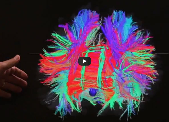
episode209However, modern neuroimaging now includes such techniques as Diffusion Tensor Imaging (DTI) and brain volume studies. The important study done by Marchi, et. al. at the Cleveland Clinic and Rochester University found abnormalities on DTI even in football players who had significant sub-concussive blows. (Subconcussive meaning less than sufficient to cause independent concussions.) Thus, if you are an athlete doing due diligence on your own brain, have DTI imaging done. For Marchi study, click here: http://www.plosone.org/article/info%3Adoi%2F10.1371%2Fjournal.pone.0056805

Brain volume studies indicate whether brain damage has resulted in the shrinkage in the size of the brain and/or substructures of the brain. Damage to important structures like the hippocampus (the brain’s save button) can now be measured. If the hippocampus is shrinking, might be a good time to stop playing.

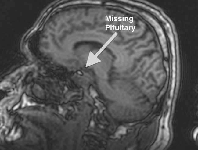
Another area which can be measured today is abnormal hormonal levels. Long term exposure to concussion can result in damage to the bodies hormonal system (endocrine system), particularly to structures such as the pituitary gland. MRI may show atrophy there and hormonal tests can also indicate pathology.

Pituitary gland damaged by trauma

The lighter spot in the center of this image is a cavity where the pituitary gland is supposed to be. Most of this pituitary gland was lost as a result of a mild traumatic brain injury.

The other approach to making return versus retire decisions, is to think of the decision in terms of reserves. Our brain’s are believed to develop with certain reserve capacity. Some of that reserve capacity is lost as we age. More is lost by traumatic events, disease, heart problems, drugs or alcohol use. Under this theory, if the amount of the loss of reserves is above the line, then perhaps there will be no disability. If the amount of reserves goes below the line, then disability will begin to show up. Think of it like you might the 53 man roster in football. A team can play with 50 players, but if the loss drops to something below 40, it might become challenging to field a team. If like the brain, there is no way to add or sign new players, if you keep having injuries, at some point there are not enough players to compete.

Demonstrated here is the concept of decreasing capacity to absorb neuron loss as an individual ages.

Demonstrated here is the concept of decreasing capacity to absorb neuron loss as an individual ages.

Age. As shown here, the older a person, the fewer reserves available. But age is more than just a matter of diminishing reserves. Age is also important because as a person approaches the age of 40, the ability of the brain to recover through a process called neuroplasticity is diminished. It is believed that neuroplasticity is closing connected to the growth hormone in the brain. The brain’s production of growth hormone drops off dramatically at age 40. The closer an athlete is to 40 when injured, the less likelihood that the brain can recover from the effects of a concussion.

No athlete wants to ever admit his or her vulnerability. No athlete wants to be forced to retire, or even miss a substantial period of time due to a concussion. Yet, while an athletic person may recover quickly from the first concussion, each successive concussion likely makes the recovery more complicated, and the potential for long term deficits more likely. While we cannot eliminate the risks of concussion in football, we can limit the disability resulting therefrom if we make sure the vulnerable minds don’t continue to be exposed.