


DTI – Diffusion Tensor Imaging MRI for Abnormal Axonal Tracts
DTI – Diffusion Tensor Imaging MRI for Visualizing Damaged Axonal Tracts
Call me at 800-992-9447
DTI is the most dynamic change in MRI technology for diagnosing those with mild traumatic brain injury. DTI is short for “diffusion tensor imaging.” DTI is a technique that visualizes axonal tracts within the white matter of the brain. Axonal tracts also called “fiber tracts“. Click here for our treatment of axonal tracts. The axonal tracts can be seen even though individual axons are too small to be seen without a microscope. Even though individual axons are microscopic, they tend to run in pathways with other axons, making a configuration that is large enough to image on DTI. As discussed in our pages on the normal brain, the Axonal tracts are similar to a large phone cable, as opposed to a telephone wire. Inside the large cable will be numerous small wires. The large cable versus the individual phone wires, is analogous to the axon tracts within the white matter of the brain.
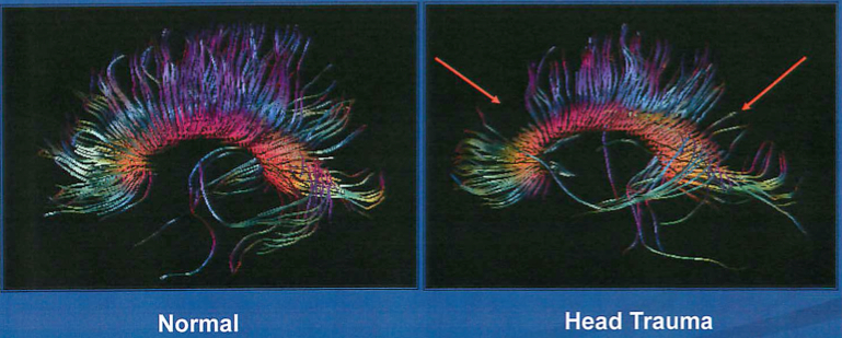
DTI can visualize the axonal tracts within the brain. Shown here are the axonal tracts of the Corpus Callosum, the axonal connections between the two hemispheres of the brain. Shown on the left is a normal brain. On the is a brain with significant traumatic loss of axonal tracts of the Corpus Callosum.
DTI is an MRI technique that is able to identify the flow of water molecules within the brain. Water molecules tend to conglomerate and move along the contours of the axonal tracts, almost as if they were a handful of straws. If you were to get several pieces of string wet and then hold them vertically, you would probably see the water running down the strand. This is essentially what can be imaged in DTI.
When diffuse axonal injury occurs in concentrated areas in the brain, we are able to see an interruption in the flow of water molecules along these fiber tracts. DTI can image this disruption, even though we cannot see any of the individual axons that are damaged. While DTI is certainly not showing all of the axonal damage throughout the brain, it is one more tool to show pathology. Considerable research on DTI has been done over the last decade, almost all of which has shown that DTI can be used for the diagnosis of permanent brain injury following concussion. An abnormal DTI scan is a strongly correlated with those concussions which cause persisting and permanent deficits. See for example – Kraus MF,White matter integrity and cognition in chronic traumatic brain injury: a diffusion tensor imaging study, Brain. 2007 Oct;130(Pt 10):2508-19. Epub 2007 Sep 14:
DTI provides an objective means for determining the relationship of cognitive deficits to TBI, even in cases where the injury was sustained years prior to the evaluation.