


3T MRI – Increased Field Strength to Image MTBI
3T MRI – 3 Tesla MRI Offers Increased Field Strength
Call me at 800-992-9447
3T MRI is short for a 3 Tesla MRI. Tesla is the measurement of the strength of a magnet. 1.5 Tesla (1.5T) is the current most common field strength of MRI scanners found in US hospitals. This is true, even a decade after 3T MRI went online for clinical use. See http://uclue.com/?xq=6379 where it is estimated that 3T MRI make up only 10% of the MRI’s scanners in the United States, as of 2013. Fortunately, most states will have access to a 3T MRI at a major hospital or university medical centers. With 550 of such machines in use, there should be a reasonable chance that someone with a persisting post-concussion syndrome, can get a referral for an MRI on 3T MRI.
One way to conceptualize the improvement in scanners is to compare such to similar improvements in the mega pixel capacity of a digital camera. An 8 mega pixel camera has roughly twice the resolution of a 4 mega pixel camera. While the difference in 3T MRI scanners from a 1.5 T MRI, isn’t exactly proportional, the analogy is valid. MRI scanners are essentially cameras that use as the contrast agent, the vibrations of magnetized protons, instead of light. An MRI could have been called a magnograph, had we given it a simple name like a photograph. In the same way that things are far clearer on a High Definition TV, the images are far clearer on a 3T MRI. What that means is that the MRI can detect smaller pathologies. The type of abnormalities that can be suspected in the brain after a concussion, are the kind of abnormalities that will be seen far better on a 3T MRI than a 1.5T MRI.
Recommendation:
- Get all Post-Concussion imaging done on a 3T MRI.
- Never use an open MRI for MTBI.
The quality of an open MRI is useless in imaging someone with a post-concussion syndrome. This would be like watching TV on a non-HD 19″ television. Even moderate to severe pathology can be missed on an open MRI, months after an injury. The below scan is of an individual who had what was called a “normal scan”, on an open MRI. On a 3T MRI, the pathology was clear:
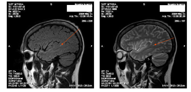
This 3 T MRI image shows (red arrow) a lesion in the brain of someone who had an open MRI, called normal.
Increased field strength is only part of potential improvement in of a 3T MRI and modern software ad ons. As more and more pathology is seen on these scans, neuroradiologists are realizing that what were considered to be insignificant findings on lower field scans, are of the pattern and nature most likely explained by traumatic forces, not disease processes or normal variants.
MRI Tailored Protocols for Identifying Pathology in Post Concussion Syndrome
The difference between a radiologist’s “call” of a “normal” versus “abnormal” may also be dependent on the protocols that a given center is using with a 3T MRI. If the neurologist or other treating physicians prescribes a “tailored protocol,” the 3T MRI can become an even more powerful diagnostic tool. A “tailored protocol“, is a set of MRI sequences, where the sequences are specific to the condition which is being be diagnosed. There is a much greater probability of finding an abnormality related to any condition if you direct the 3T MRI towards what it is you are looking for. If you find something, it is worth the effort. (Of course corporations and insurance companies will always whine about any extra money or effort to identify any pathology.)
If the radiologist is not directed to seek subtle pathology on 3T MRI, he or she may not see it. If the same techniques and protocols are used as if the search was for acute hemorrhage, the chances of a scan finding subtle yet significant pathology go down exponentially. The burden of such a common sense approach to a 3T MRI, should lie with the neurologist who prescribes the scan. But as not all neurologist understand the potential of a 3T MRI, you might want to ask for the additional protocols we suggest on the next pages.